#0065 Radiographic diagnosis of Pneumoconioses by AIR Pneumo-trained physicians: Comparison with low-dose thin-slice computed tomography
Comparing Screening Technologies and the Diagnosis of Lung Conditions in Construction Workers
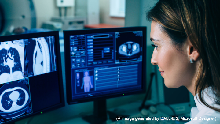
Pneumoconiosis is a lung condition that typically affects people like miners and construction workers who breathe in a large amount of dust particles as they go about their work. Chest radiography can easily pick up this condition, and training programs such as the Asian Intensive Reader of Pneumoconiosis (AIR Pneumo) and the US-based National Institute of Occupational Safety and Health’s (NIOSH) B reader certification help physicians improve their diagnostic skills in interpreting chest radiographs accurately. Computed tomography (CT), a technique that takes x-ray scans of organs and bones from different directions to produce several images in thin slices, has demonstrated greater accuracy and sensitivity in detecting the early stages of the disease.
To judge how well thin-slice CT scans and chest x-rays compared in picking up lung diseases through occupational exposure, we assessed the thin-slice CT scans and chest x-rays of nearly 100 construction workers, also checked whether physicians trained in AIR Pneumo and NIOSH B could successfully identify such diseases in both mediums.
On assessing the chest x-rays, we found that AIR Pneumo and NIOSH B clinicians were highly sensitive and specific at detecting benign asbestos-related diseases as compared to thin-slice CT results. Small plaques, caused by the build-up of chalky material in the chest wall and diaphragm on exposure to asbestos, were much more easily detectable on thin-slice CT but not on chest x-rays. We also observed inaccuracies such as false positives and false negatives in chest x-rays, as well as rib fractures being wrongly identified as pleural plaques in some cases.
The study highlighted the importance that years of experience can make in diagnostic performance, with physicians who had three years of experience and were trained through the AIR Pneumo program performing on par with those trained in the NIOSH B program. It is a testament to the improvements in working conditions that there was a noted absence of severe pneumoconiosis symptoms on both thin-slice CT images and chest x-rays.
Our research demonstrated that thin-slice CT is excellent at detecting disease characteristics that perhaps a regular chest x-ray may not pick up. However, chest radiographies are an affordable tool to screen several employees for lung conditions. An experienced physician with a heightened diagnostic sensitivity can use them to effectively assess workers’ health.
Link to the original journal article:
https://onlinelibrary.wiley.com/doi/10.1002/1348-9585.12141
Title of the paper:
Radiographic diagnosis of Pneumoconioses by AIR Pneumo-trained physicians: Comparison with low-dose thin-slice computed tomography
Authors:
Shoko Nogami, Naw Awn J-P, Munenobu Nogami, Tomomi Matsui, Nlandu Roger Ngatu, Taro Tamura, Yukinori Kusaka, Harumi Itoh, Narufumi Suganuma